


Optical imaging and analysis systems for fluorescent, bioluminescent in vivo signals (laboratory animals) BIOVIVO™ B series (Wuhan BIOVIVO Biotechnology Co., Ltd., China)
Wuhan BIOVIVO Biotechnology Co., Ltd. is a high-tech company specializing in the development, manufacturing, sales, and technical support of in vivo imaging systems (for plants and animals).
The company’s core business is the manufacture of equipment and the development of technologies for the in vivo molecular imaging of living organisms. By integrating related technologies in the field of plant and animal imaging, the company manufactures equipment for a wide range of biotechnology projects, including plant genetic transformation, cell genome editing, and in vivo molecular imaging of living organisms.
In vivo optical imaging technology is used to track, screen, and quantitatively analyze bioluminescent or fluorescent markers in living organisms. Applications span fundamental research, including genetic research, oncology, stem cell research, and disease studies, as well as preclinical drug evaluation and screening.
Based on market needs and drawing on extensive experience in in vivo imaging technologies, the company analyzed and addressed key challenges in the in vivo imaging equipment market. Based on this, the company independently developed a new generation of BIOVIVO™ series in vivo imaging systems, protected by independent intellectual property rights. The series includes the BIOVIVO™ B animal imaging systems and the BIOVIVO™ G plant imaging systems. These systems offer optimal solutions for bioluminescent and fluorescent imaging of laboratory animals and plants, ensuring convenient, fast, and efficient in vivo data collection and analysis.


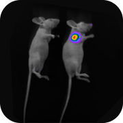


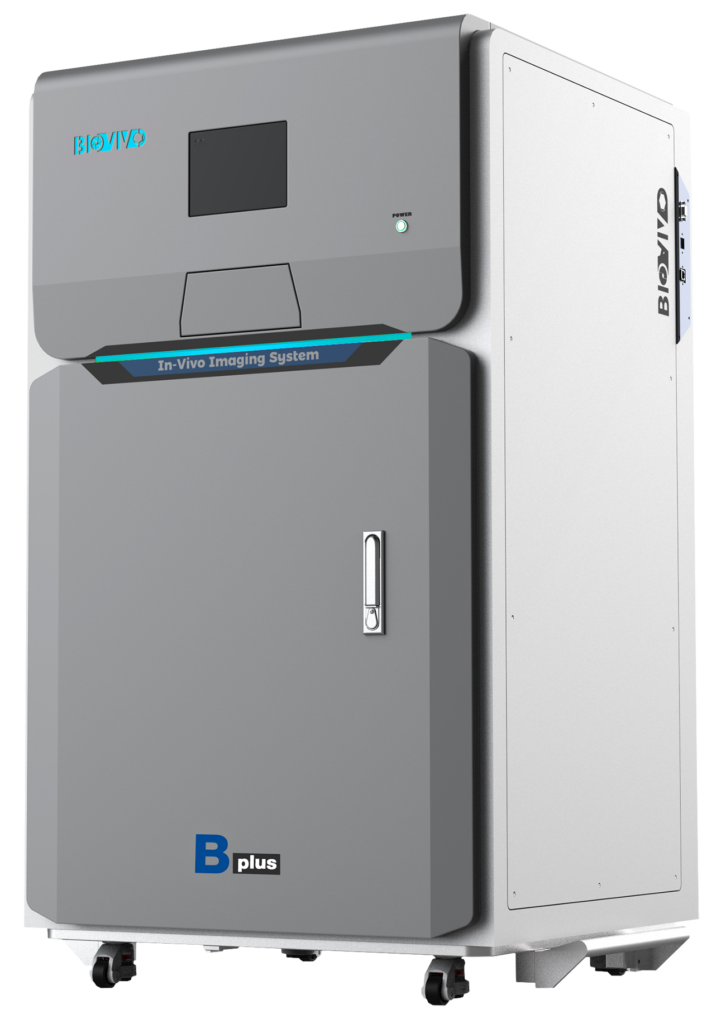



As early as the first two decades of the 20th century, and even earlier in the 19th century, scientists discovered that focusing on relatively simple organisms could partially unravel the mysteries of developmental phenomena. For example, in uncovering the laws of heredity in nature, Mendel used peas as experimental material, while Morgan chose the fruit fly Drosophila. In their studies, peas and Drosophila had fewer cells, their structure was more homogeneous, and changes were easier to observe.
As a result of evolution, cellular life forms exhibit significant similarities in fundamental developmental patterns, and many basic life mechanisms are similar across various biological species on Earth. Therefore, it is possible to use species at relatively low steps of the biological complexity ladder to study general developmental patterns. This is especially true when common characteristics of morphogenesis and changes are found in organisms with different developmental features, allowing for the establishment of universal principles of development. Studying such organisms helps us uncover universal patterns of life phenomena and understand the general laws governing the living world. Thus, these specially selected organisms were named “model organisms.” The choice of a specific organism as a model depends primarily on the scientific task the researcher sets, as well as on finding the species most suitable for solving this task.
In the early 20th century, Morgan chose the fruit fly Drosophila melanogaster as his research subject. Based on this work, the chromosomal theory of heredity was established, laying the foundation for classical genetics and initiating the use of Drosophila as a model organism. Scientists not only confirmed Mendel’s laws in Drosophila but also discovered sex-linked inheritance of the white-eye mutation, formulated the principle of linear gene arrangement on chromosomes, and the law of linkage and crossing over. In 1933, Morgan was awarded the Nobel Prize for these achievements. The sequencing of the Drosophila genome, completed in 2000, solidified its status as a crucial model animal for studying genetics and developmental biology.
Since 1965, scientist Sydney Brenner introduced the nematode Caenorhabditis elegans into the field of molecular and developmental biology research. In 1983, the complete cell lineage of the worm from fertilized egg to adult was described, marking a milestone in the history of developmental biology. Subsequently, C. elegans found widespread application in studies of embryonic development, sex determination, apoptosis, behavior, and neurobiology, and became a crucial model organism in the study of aging and lifespan.
Zebrafish possess characteristics such as high fecundity, external fertilization and development, transparent embryos, a short sexual maturity cycle, small size, and ease of maintenance, making them an ideal choice for studying mechanisms of embryonic development, gene functions, and disease pathogenesis. The African clawed frog (Xenopus laevis) has large and abundant oocytes, which are convenient for microsurgical manipulations. Furthermore, they can be used to create biologically active cell-free systems suitable for biochemical analysis, ensuring their indispensable role in developmental biology research.
The main model animal is, undoubtedly, the laboratory mouse! Due to their small size, docile nature, ease of maintenance, and the similarity of developmental processes and anatomical tissue structure to humans, mice began to be used in anatomy and animal experiments as early as the 17th century. As a result of long-term selection and breeding under artificial conditions, thousands of independent outbred and inbred lines have been developed to date. The laboratory mouse has become the most important model organism for deciphering human gene functions and studying human diseases, as well as the most thoroughly studied mammal in the world for scientific experiments. Moreover, mouse genes exhibit a high degree of homology with human genes: 99% of human genes have corresponding genes in the mouse genome. The mouse is widely used as a model organism in biomedical research and is the most detailed studied mammal for experiments in the modern world.
Above were presented model organisms from the animal kingdom, which we commonly refer to as model animals. Are there also model plants? The answer is affirmative. In modern botanical research, Arabidopsis thaliana, tobacco, tomato, rice, and others are most frequently used as model plants, with each serving its specific research purposes.
Arabidopsis thaliana opens the list of model plants. It belongs to the division of angiosperms, the class of dicotyledons, and is a representative of the mustard family. This plant itself does not possess great economic value. However, its small size, high seed productivity, and short life cycle; clearly distinguishable morphological features, convenient mutant phenotypes for observation, and self-pollination make it an exceptionally convenient object. The genome of Arabidopsis thaliana is considered small for higher plants (about 125 million base pairs and 5 pairs of chromosomes), and its genes are characterized by a high degree of homozygosity. Treatment with physicochemical factors leads to a high frequency of mutations, making it easy to obtain mutants with defects in various metabolic functions. Thanks to this, Arabidopsis thaliana has become an ideal model organism for studying the genetics, cellular development, and molecular biology of flowering plants, earning it the title of “Drosophila of the plant world” among scientists.
The history of Arabidopsis thaliana research dates back to the 16th century. In 1943, Laibach thoroughly substantiated its advantages as a model organism, which contributed to the first International Conference on Arabidopsis thaliana in Germany in 1965. However, its active use as a model organism began approximately in the last two decades of that century. In 1986, the Meyerowitz laboratory first reported the cloning of an Arabidopsis thaliana gene. In 1988, the first RFLP map of its genome was published. In the following few years, reports of gene cloning of mutants with T-DNA insertions and gene cloning based on genetic maps followed. In 2000, the work on determining the complete nucleotide sequence of its genome was completed, making Arabidopsis thaliana the first plant with a fully sequenced genome.
Tomato, or love apple, is an annual or perennial herbaceous plant of the nightshade family, genus Solanum. The plant height is 0.6–2 meters, its entire surface is covered with sticky glandular hairs, and it has a strong characteristic odor. The stem tends to lodge. Leaves are pinnately compound or pinnately dissected. The peduncle length is 2–5 cm, the inflorescence usually consists of 3–7 flowers. The calyx and corolla are wheel-shaped. The fruit is a juicy fleshy berry, oblate-spherical or almost spherical, with yellow seeds. The flowering and fruiting period occurs in summer and autumn. Due to its relatively small genome size, short life cycle, ability to self-pollinate, well-established genetic transformation system, and clear genetic basis, as well as a more distant relationship with other model plants (such as Arabidopsis thaliana or rice), tomato has become a classic model organism for studying fruit development and ripening processes.
Rice, as one of the most important cereal crops in the world, plays an increasingly significant role as a model plant for studying cereals. This is due to its relatively small genome, known genomic sequence, well-established Agrobacterium-mediated genetic transformation system, and high degree of homology (genetic similarity) with other cereal crops. Tobacco is not only an important economic crop but also one of the most valuable materials for scientific research. Many innovative studies have been conducted on tobacco. A significant portion of botanical knowledge, such as photoperiodism, plant nutrition, photosynthesis, photorespiration, organic metabolism, as well as virus and transgenic organism research, has been obtained from tobacco. No other plant has been studied as deeply by science as tobacco. It is a model organism for molecular biology and genetic engineering and has rightfully earned the title of “laboratory mouse of the plant kingdom.”
In addition to the aforementioned model animals and plants, the family of model organisms, of course, also includes microorganisms. Escherichia coli and yeast are key materials for studying the genetics of microorganisms and are widely used as model organisms in microbiological research. In the future, as research in life sciences develops, the family of model organisms will undoubtedly continue to grow.
Key stages of an in vivo optical imaging experiment in mice
In vivo optical imaging in laboratory animals, particularly mice, primarily relies on two technologies: bioluminescence and fluorescence. Bioluminescence involves labeling cells or DNA with the Luciferase gene, while fluorescent technology uses fluorescent reporter genes (e.g., green fluorescent protein, red fluorescent protein), fluorophores (such as FITC, Cy5, Cy7), and quantum dots (QD) for labeling.
- Preparation of laboratory animals: selection of a mouse line appropriate for the experimental objectives, performing necessary acclimatization and adaptation to housing conditions.
- Introduction of a fluorescent probe or bioluminescent label: according to the research objectives, a suitable fluorescent probe or bioluminescent marker is selected and administered to the experimental animal.
- Pre-imaging preparation: before starting the imaging procedure, the animal must be prepared, such as fur cleaning and fixation in the required position, to ensure accuracy and reproducibility of results.
- Imaging procedure: depending on the chosen in vivo imaging technology, the labeled animal is placed in the appropriate imaging equipment. During the procedure, environmental parameters (temperature, humidity, etc.) must be controlled to ensure image quality and adherence to bioethical principles.
- Data analysis and interpretation: after image acquisition, data analysis is performed. Results are evaluated and interpreted in the context of the experimental design and research objectives.
Key labeling methods in in vivo optical imaging technology in laboratory animals
| Bioluminescence | 1. Luciferase gene labeling: | The Luciferase gene is used to label cells or DNA. |
| Fluorescence | 1. Fluorescent protein labeling: | Fluorescent proteins are suitable for labeling cells, viruses, genes, etc. GFP, EGFP, RFP (DsRed) are most commonly used. |
| 2. Fluorescent dye labeling: | The principle of fluorescent dye labeling is similar to in vitro labeling methods. Cy3, Cy5, Cy5.5, and Cy7 are frequently used to label antibodies, polypeptides, micromolecular drug compounds, etc. | |
| 3. Quantum dot labeling: | Quantum dots, as a new class of fluorescent labels, offer unique advantages for long-term monitoring of biological processes and in vivo tracking. |
Principle of in vivo optical imaging in mice
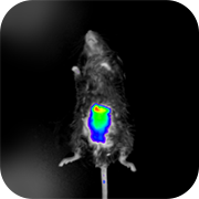
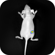
In vivo imaging technology for small laboratory animals is based on the use of a high-sensitivity cooled CCD camera combined with a specialized light-tight chamber and image processing software. This allows for direct observation of cell activity and gene expression in a living organism. By recording data from the same group of experimental subjects at different time points, the movements and changes of a single observation target (labeled cells and genes) are tracked. The method is characterized by ease of execution, clear results, and high sensitivity, and is widely applied in various fields, including life sciences, medical research, and drug development. In oncology: tumor models allow in vivo imaging to measure tumor growth and metastasis, as well as its response to drugs, bringing oncology research closer to the microenvironment of clinical disease. Compared to traditional methods, this approach not only increases sensitivity (allowing detection of microscopic tumor foci) but is also suitable for quantitative analysis of in vivo tumor growth, eliminating inter-individual differences that arise when animals need to be sacrificed for analysis. In drug research: in vivo imaging can be used to study genes related to drug metabolism, their mechanisms of action, as well as target organs and drug distribution patterns in the body.

In vivo optical imaging technology has become a powerful tool in both fundamental biomedical research and medical diagnostics. By tracking and recording data from the same group of subjects at different time points, this technology facilitates a more convenient and effective understanding of the patterns of human disease onset and development, as well as research into methods of prevention and treatment. This technology is already widely used in areas such as oncology, pharmacological research, gene therapy, apoptosis studies, and epidemiology.

Fig. 1. Principle of luminescence in in vivo imaging |
Figure 1. Use of Luciferase for labeling genes, cells, and living organisms. Luciferase, a protein expressed by the luciferase gene, and the fluorescein substrate, in the presence of oxygen, Mg²⁺, and ATP, undergo an oxidation reaction, converting part of the chemical energy into light. This luminescence is recorded externally by a high-sensitivity CCD camera to form an image. A single injection of fluorescein into a laboratory mouse allows the luminescence of luciferase-labeled cells to be maintained in its body for 30-45 minutes.
- Pharmacodynamic evaluation of anti-tumor drugs in vivo using an oncolytic virus model. Newcastle disease virus (NDV) has been used in oncolytic therapy for decades due to its natural oncolytic properties. α2,6-linked sialic acid plays a key role in NDV binding and infection of tumor cells. In an article published by Li, Q. et al. in 2017 [1], NDV was labeled with luciferase. In vivo imaging experiments demonstrated that NDV has a pronounced anti-tumor effect against SW620 colorectal cancer cells with high expression of α2,6-sialic acid on the cell surface, while its effect on SW480 cells with normal expression levels of α2,6-sialic acid was insignificant.
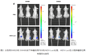
Fig. 2. After subcutaneous implantation of SW620 and SW480 tumor cells into mice, treatment with PBS and rNDV-Luci was performed. In the rNDV-Luci treated group, an intense fluorescent signal was observed in the tumor area. |
- Studies on the mechanism of tumor growth suppression by gene silencing have shown that COPB2 is significantly overexpressed in gastric cancer cell lines. In the work by An, C. et al. [2], it was demonstrated that COPB2 knockout leads to the suppression of the RTK signaling pathway and its downstream cascade molecules, which promotes apoptosis of gastric cancer cells and inhibits tumor growth in nude mice. The results of in vivo imaging experiments showed that the total fluorescence intensity in mice infected with Lv-shCOPB2 was significantly lower compared to the control group infected with Lv-shRNA.
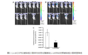 Fig. 3. The total fluorescence intensity in mice infected with Lv-shCOPB2 is significantly lower compared to the control group infected with Lv-shRNA. Fig. 3. The total fluorescence intensity in mice infected with Lv-shCOPB2 is significantly lower compared to the control group infected with Lv-shRNA. |
- Searching for new therapeutic targets for cancer treatment: A research group from the School of Pharmacy, Fudan University, in an article published in 2017 [3], showed that TIPE2 overexpression significantly suppresses the proliferation of 4T1 cells both in vitro and in vivo. TIPE2 increases the number of T cells and NK cells and reduces MDSCs. TIPE2 enhances the production of IFN-γ and TNF-α by CD8⁺ T cells and NK cells in the tumor microenvironment, boosting their cytotoxic activity. The suppression of TIPE2 in breast cancer development and metastasis is likely mediated by enhancing CD8⁺ T cell and NK cell-mediated anti-tumor immune responses. Thus, TIPE2 may be a potential therapeutic target for breast cancer treatment.
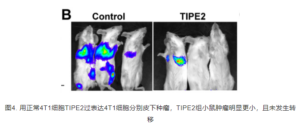
Fig. 4. Upon subcutaneous implantation of normal 4T1 cells and 4T1 cells with TIPE2 overexpression into mice, the group of animals that received cells with TIPE2 overexpression showed significantly smaller tumors and no metastases were detected. |
- Investigation of CAR-T cell therapy efficacy in solid tumors: Chimeric antigen receptor T-cell (CAR-T) therapy demonstrates limited efficacy in treating solid tumors, primarily due to their weak ability to reach and penetrate tumor sites. Structural features of solid tumors, loss of specific antigens, and a highly immunosuppressive microenvironment pose major challenges for CAR-T therapy in treating this type of neoplasm. In a study targeting head and neck squamous cell carcinoma (HNSCC), MUC1 was chosen as the target [4]. The authors constructed both a second-generation CAR and a fourth-generation CAR capable of simultaneously secreting IL-22. In vitro and in vivo experiments showed that CAR-MUC1-IL-22 T cells possess stronger and more effective cytotoxic activity against MUC1+ HNSCC cells compared to conventional CAR-T cells. These results indicate the potential efficacy of CAR-T therapy for patients with HNSCC and lay the scientific foundation for treatment using MUC1+ CAR-T cells.
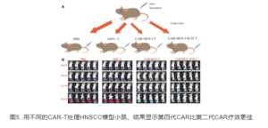
Fig. 5. When treating mice with an HNSCC model with different CAR-T cell variants, results showed that the fourth-generation CAR had higher therapeutic efficacy compared to the second-generation CAR. |
In a recent study conducted by a joint team of scientists from the Institute of Zoology, Chinese Academy of Sciences, and Peking Union Medical College Hospital, gene expression profile interaction analysis revealed that C-C chemokine ligand 20 (CCL20) has high expression in lung cancer, as well as in other cancers with high incidence and/or mortality, such as colon cancer, rectal cancer, gastric cancer, and liver cancer [5]. Forced expression of C-C chemokine receptor type 6 (CCR6) led to the migration of CAR-T cells to tumor cells secreting CCL20. In a lung cancer xenograft mouse model, CAR-T cells with CCR6 overexpression, after administration, effectively migrated and infiltrated the solid tumor, leading to effective tumor elimination and a significant increase in mouse survival. The results of this study provide supporting data for the clinical development of CAR-T cells with engineered chemokine receptors in solid tumor immunotherapy.
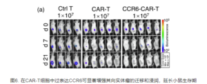
Fig. 6. CCR6 overexpression in CAR-T cells significantly enhances their migration and infiltration into solid tumors, leading to increased mouse survival. |
References
【1】 Li, Q., Wei, D., Feng,F. et al. α2,6-linked sialic acid serves as a high-affinity receptorfor cancer oncolytic virotherapy with Newcastle disease virus. J CancerRes Clin Oncol 143, 2171–2181 (2017).
【2】 An, C., Li, H., Zhang, X.,Wang, J., Qiang, Y., Ye, X., Li, Q., Guan, Q., Zhou, Y.”Silencing of COPB2inhibits the proliferation of gastric cancer cells and induces apoptosis viasuppression of the RTK signaling pathway”. International Journal ofOncology 54.4 (2019): 1195-1208.
【3】 Zhenhua Zhang, Li Liu,Shousong Cao, Yizhun Zhu, Qibing Mei, Gene delivery of TIPE2 inhibits breastcancer development and metastasis via CD8+ T and NK cell-mediated antitumorresponses, Molecular Immunology, Volume 85, 2017, Pages 230-237, ISSN0161-5890.
【4】 Mei, Z, Zhang,K, Lam, AK‐Y, et al. MUC1 as a target for CAR‐T therapy in head and neck squamous cell carinoma. CancerMed. 2020; 9: 640– 652.
The information about BIOVIVO™ equipment provided on this website is based on standard technical specifications and is for informational purposes only. The exact technical specifications and configuration of BIOVIVO™ equipment are determined by a specific engineering solution designed to meet the customer’s needs!


